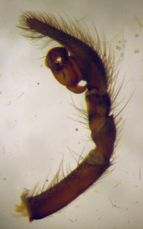
prepared left palpus of a male specimen of Pireneitega spasskyi (Amaurobiidae)
In arachnology the most common preparations to be made are mere “amputations” of body parts to be examined separately within the ethanol solution (ω = 70-80 %) which is also used for conserving whole spider specimens. Sometimes this helps when counting and measuring leg bristles or tarsal claws but most often the higly specialized and species-specific pedipalps of mature male specimens are prepared in this way (e. g. torn off) for close examination in the process of determining which species this specimen belongs to.
The other important preparation is the preparation and clearing (see details) of the female epigyne in order to examine the structure of the inner genital organs (e.g. vulva). In a first step the visible epigyne is cut out generously, taking caution not to damage the sclerotized epigynal parts nor the structures, which are not visible from the outside. Also, the spider body should not be damaged in the process. This takes some practice, especially when the spider itself is only 2-3 mm in size! This “cutting out” of the epigyne is not actually done with scissors but most conveniently with two pairs of thin stainless steel forceps.

preparing an epigyne/vulva. white forceps holding the spider, black forceps slowly tearing cuticula around epigyne (Pireneitega spasskyi, Amaurobiidae).
One pair of forceps firmly holds the spider in position (venter facing upwards) by gripping the spider next to the epigyne at the epigastric fold, while the second pair of especially thin and pointy forceps is used to tear open the cuticula directly next to the first pair of forceps – thus tearing open the ventral cutucula starting at the epigastric fold and proceeding in a semicircle around the epigyne with one pair of forceps holding the cuticula near the torn cuticula and the thinner pair of forceps tearing the cuticula somewhat more.
After the epigyne has been cut out, it can then be put onto a microscopic slide and covered with a cover slip. The ethanol solution can be used as a (very!) short-term mounting fluid but will evaporate within a few minutes even if covered with a cover slip. This is a clear disadvantage of ethanol solution as a mounting fluid, next to the fact that soft tissues (cartilage, muscle) attached to genital structures of interest do obscure the view, thus complicating observations. More or less permanent preparations and/or effective dissolving of soft tissues can be done using special clearing and mounting media, e. g. the famous Hoyer’s mounting medium (details here).
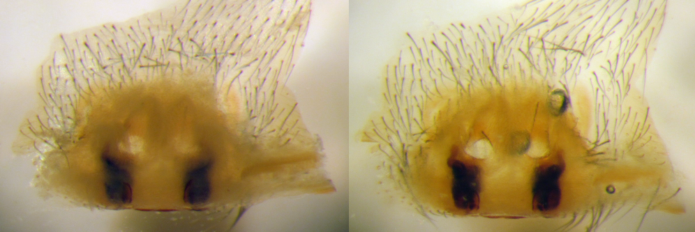
slide-prepared vulva of Pireneitega spasskyi (Amaurobiidae): left – in ethanol, right: in Hoyer’s solution. Notice the much better visible structures in the right photo.

storing prepared body parts in a small plastic tube makes them easily discernible in the vial
Thus prepared epigynes and also small palps should always be stored together with the spider for later referral. They can however, easily get lost when floating around seperately in a vial. Male palps can sometimes be fixed between two legs of the spider but a better method is to put the palp (or an epigyne) in a small plastic tube and close it on both sides with cotton balls. Such a tube cannot get lost when stored in a vial together with the spider and the location label.
Erst einmal vielen Dank für die Methode!
Vlt. kann man noch ergänzen, dass es bei Weibchen größerer Spinnen (da gehören viele Amaurobiidae auch dazu) immer lohnend ist mit Präpariernadeln/Minutien zu versuchen, das Zwischengewebe um die Vulva herum zu entfernen. Desto klarer wird dann auch die aufgehellete Ansicht (z.B. nach Anwendung mit Hoyers Medium).
Und noch eines: Die Härchen um die Epigyne herum (nur, falls diese die Sicht auf wichtige Strukturen verdecken/erschweren sollten) lassen sich mi einer geschliffenen Pinzette meistens recht gut abschaben. Beim Abschaben sollte man die Epigyne noch nicht herauspräpariert haben, denn aufgrund des Flüssigkeitsinnendruckes des Opisthosomas (das man dabei leicht zusammendrückt) lässt sich sehr gut arbeiten und eine Beschädigung der umgebenden Kutikula oder gar der Epigyne selbst vermeiden.
Viele Grüße
Steffen
Danke Steffen für die Hinweise. Genauso mache ich das auch: Erst physikalisch mit Pinzette & Co. vorbereiten und dann chemisch mit Hoyers Lösung aktiv werden.